FACTS Art of Science 2023
Consolation Prizes
Fatigue crack-arrest by dynamic recrystallization (DRX)

Submitted by: Jayaraj Radhakrishnan, Prof. Upadrasta Ramamurty (Supervisor)
School/Centre: MAE
Model of Equipment: FESEM 7800F
Description: BSE/EBSD was employed to find DRX in the vicinity of lack of fusion pores of IN718 fabricated by laser beam powder bed fusion and subjected to cyclic loading at 600degC. After crack initiation, crack-path deviated around these newly nucleated fine grains (which were otherwise absent before test) and increased fatigue resistance. This could improve the life of turbine blades and delay failure in aeroengines.
The inverse pole figure of Fe-3.5wt%Si@SiO2 nanocomposite fabricated via laser powder bed fusion

Submitted by: Xiaojun Shen, Assist Prof Christopher H.T. Lee (Supervisor)
School/Centre: EEE
Model of Equipment: FESEM 7800F
Description: This image is acquired by an EBSD detector (symmetry, Oxford instrument, UK) equipped on a Field Emission Scanning Electron Microscopy (FESM, JEOL Model 7800, Japan). By adding high resistivity SiO2, Fe-3.5wt%Si@SiO2 was additively manufactured via laser powder bed fusion technique for electric motor application to reduce eddy current loss.
EBSD of helicoidal microstructure under compressive deformation

Submitted by: Crystal Ji, Prof Bartolo (Supervisor)
School/Centre: MAE/SC3DP
Model of Equipment: FESEM 7800F
Description: The sample was made by laser powder bed fusion technique. By modifying the scan strategy, we are able to achieve this gradient material with distinctive microstructure. This image is captured by EBSD, after sample has undergo 30% strain compression.
Hairs on a bee's wing

Submitted by: Quentin Perrin, Prof Ali Miserez (Supervisor)
School/Centre: MSE
Model of Equipment: FESEM 6340F
Description: LEI imaging of a bee's wing. This illustrates the Salvinia effect in nature, keeping a permanent layer of air between the microscopic hair for better hydrophobicity and aerodynamics. Biomimetic applications of this effect reduce drag in cargo ships, reducing fuel consumption. This sustainable solution can therefore avoid conventional polluting coatings.
Akin to a soap sponge on the surface but with filled pores like a layered cake

Submitted by: Nupur Gupta, Prof. Hu Xiao (Supervisor)
School/Centre: MSE
Model of Equipment: FESEM 7600F
Description: The image represents a cross-section of the poly N-isopropyl acrylamide/ poly sodium acrylate semi-IPN hydrogel with a polyamide layer on its surface (https://pubs.acs.org.remotexs.ntu.edu.sg/doi/full/10.1021/acsami.1c16639). The hydrogel sample was lyophilized and sputter coated before observing under FESEM 7600 in SEI mode. The image resembles akin to a soap sponge on the surface, but with filled pores like a layered cake. The material was synthesized by me.
Cross-sectional STEM for SnS/WS2 superlattice

Submitted by: Zhou Xin, Assist Prof. Song Peng (Supervisor), Prof. Liu Zheng (Co-supervisor)
School/Centre: MSE
Model of Equipment: TEM ARM 200F
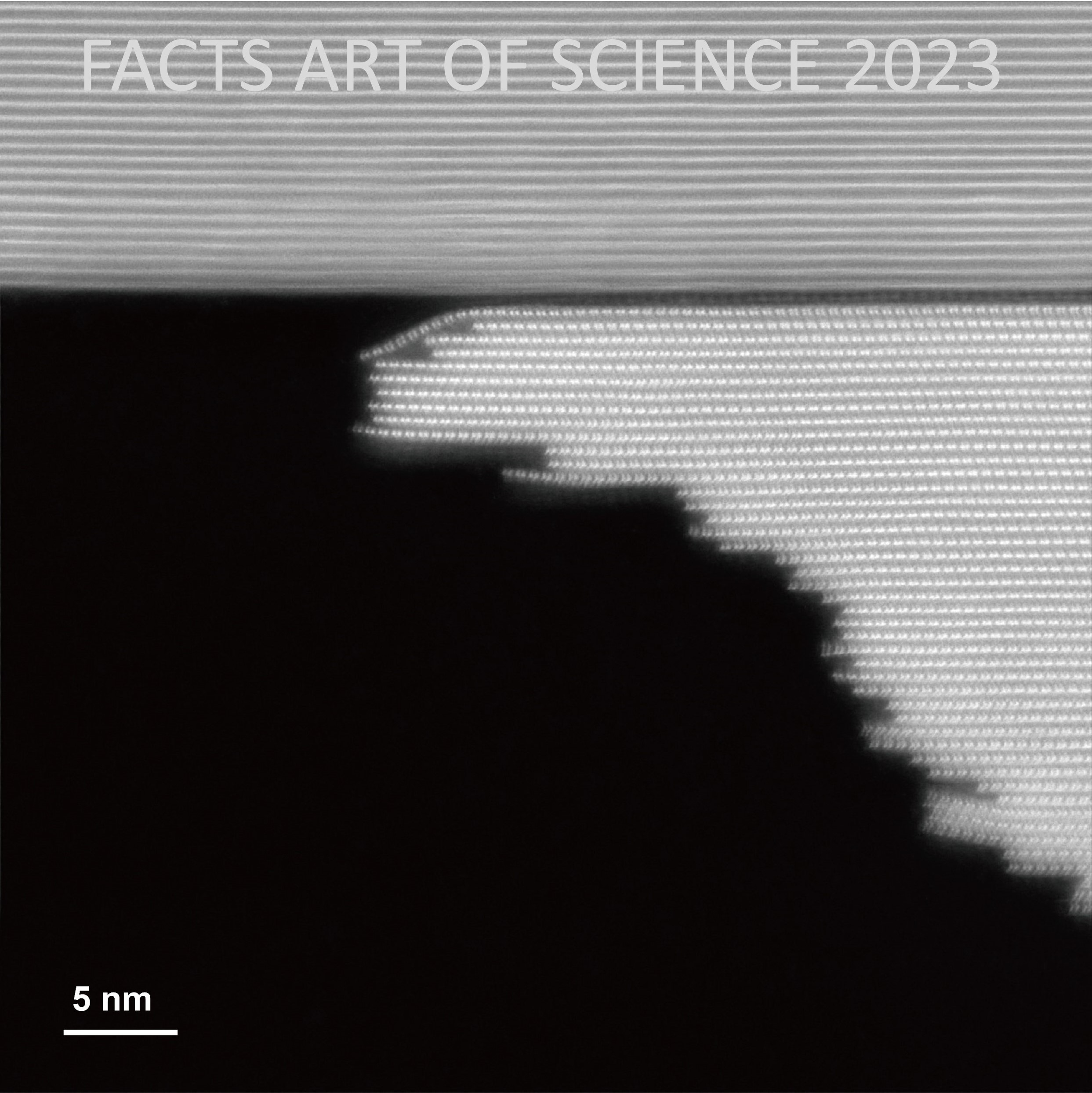
Description: Cross-sectional STEM for SnS/WS2 superlattice, by ARM200F at 200 kV. The sample was prepared and examined by Zhou Xin, from Prof. Liu Zheng and Prof. Song Peng’s group.
Tungsten Selenide Cross-section

Submitted by: Wu Yao, Assoc Prof. Li Shuzhou (Supervisor), Prof. Liu Zheng (Co-supervisor)
School/Centre: MSE
Model of Equipment: TEM ARM 200F
Description: The 2H phase tungsten selenide (WSe2) cross-section was imaged using the aberration-corrected HAADF-STEM. The intralayer abc stacking sequence and interlayer AA' sequence of W and Se atoms could be seen clearly from the image.
Twisted Molybdenum Disulfide

Submitted by: Wu Yao, Assoc Prof. Li Shuzhou (Supervisor), Prof. Liu Zheng (Co-supervisor)
School/Centre: MSE
Model of Equipment: TEM ARM 200F
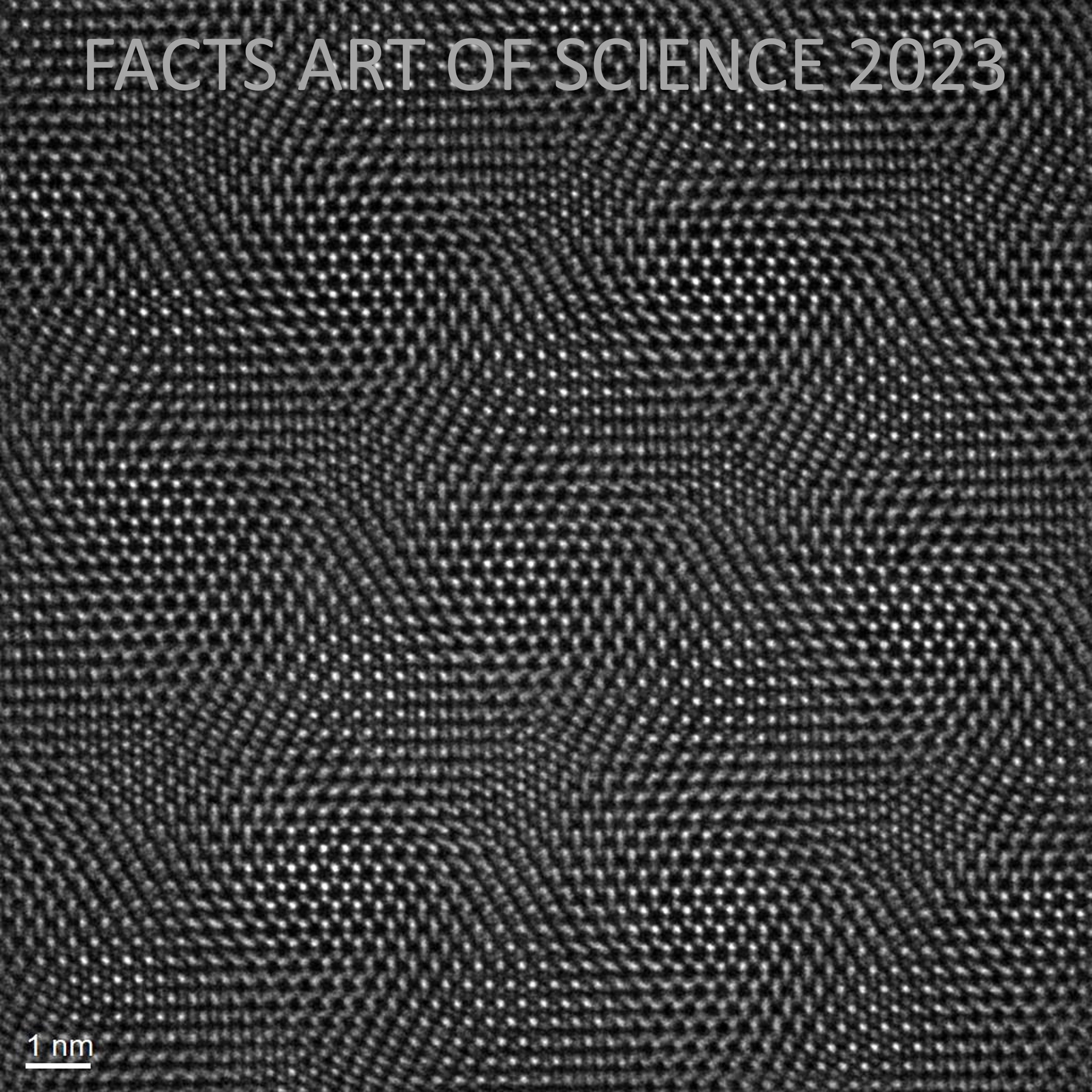
Description: Bilayer molybdenum disulfide with a twisted angle was imaged using an aberration-corrected HAADF-STEM. Due to interlayer rotation, the material exhibits significant moire superlattices. This moire superlattice has become a hot spot in semiconductor physics, potentially applied in superconductive materials and new electronic devices.
Atomic-resolution aberration-corrected HAADF-STEM image of flame-synthesized TiO2-x under 15 min calcination

Submitted by: Wu Shuyang, Dr Zhang Zhengyang (Supervisor)
School/Centre: School of Chemistry, Chemical Engineering and Biotechnology
Model of Equipment: TEM ARM 200F
Description: Blue TiO2-x with controllable defect content and location is fabricated by the one-step and solvent-free flame synthesis method. The TiO2-x with 15 min calcination time (TiO2-x-15 min) contains multiple types of defects including bulk defects, stepped surface and stacking faults. It shows remarkable photocatalytic activities in H2 generation, CO2 reduction and pollutant degradation.
h0l precession image of imidazolium lead bromide at 220 K revealing a rare 4-fold superstructure combined with threefold twinning

Submitted by: Walter Wong, Assoc Prof Andrew Grimsdale (Supervisor)
School/Centre: MSE
Model of Equipment: XRD Bruker Smart Apex II
Description: Single crystal XRD was carried out on imidazolium lead bromide at 220 K. This h0l precession image shows the presence of satellite peaks revealing a rare 4-fold superstructure. The apparent hexagonal shape is a result of threefold twinning (i.e. 3 domains at 60° apart). Sample were made by myself.
Consolation Prizes
Artificial Intelligence (AI) Robot of Molybdenum Telluride

Submitted by: DENG YA, Prof. Liu Zheng (Supervisor)
School/Centre: MSE
Model of Equipment: FESEM 7600F (TED/EBL)
Description: The Image was taken using by SEM-SEI technique, and the object is NaCl-assisted synthesized MoTe2 flakes. The bright particles in the center area are salt which is widely used for our 2d materials synthesis. This image resembles an Artificial Intelligence (AI) robot, which also indicates the possibility of AI-assisted material synthesis in the future. The sample is prepared and photographed by Deng Ya.
A blooming microscopic world

Submitted by: Ye Kai, Assoc Prof Aravind Dasari (Supervisor)
School/Centre: MSE
Model of Equipment: FESEM 7600F (TED/EBL)
Description: This is a SEI micrograph of the graphite electrode from a lithium-ion battery when exposed to atmospheric conditions. The flower-shape morphology was formed during its decomposition when exposed to moisture and oxygen in air. A beautiful ending for a beautiful material.
The Bamboo Forest

Submitted by: Rodney Chua Yong Sheng, Prof Madhavi Srinivasan (Supervisor)
School/Centre: Energy Research Institute @ NTU
Model of Equipment: FESEM 7600F
Description: This piece of “Bamboo Forest” inspired art is a scanning electron micrograph of a green cathode material deposited onto carbon fibres via the facile hydrothermal method for high-performance energy storage applications.
ZnO Nanoflower Field

Submitted by: Ronn Goei, Assoc. Prof. Alfred Tok (Supervisor)
School/Centre: MSE
Model of Equipment: FESEM 7600F
Description: ZnO nanoflower field - prepared by growing ZnO hydrothermally onto glassy carbon electrode. The ZnO will be used as a piezoelectric sensor materials for soft robotic gripper application.
Fern leaves

Submitted by: Nguyen Linh Lan, Prof. Lam Yeng Ming (Supervisor)
School/Centre: MSE
Model of Equipment: Zeiss Crossbeam X540
Description: Perovskite crystals after being spin coated and annealed on a glass substrate resembling fern leaves. The material was synthesized from one-step spin coating techniques of precursor solution consisting of equimolar MAI and PbI2. The material is expected to have favourable optoelectronic properties for LED application.
When Fall Arrives

Submitted by: Tso Shuen, Prof Alex Yan (Supervisor), Dr. Xu Jian Wei (Co-supervisor)
School/Centre: MSE
Model of Equipment: FESEM 7600F (TED/EBL)
Description: SEI from SEM 7600F was used. This image shows the forest ground when fall arrives. In the middle is antimony sulfide that is used as anode of sodium battery. It resembles as the last two petals fallen from the tree. There is a carbon nanotubes cluster at the upper part of the image as a pinecone. Surrounding graphene flakes resemble as wood chips and rocks with green moss covering their edges. This sample belongs to Zhao Wenqing.
Spider book lungs

Submitted by: Quentin Perrin, Prof Ali Miserez (Supervisor)
School/Centre: MSE
Model of Equipment: FESEM 7600F
Description: SEI imaging of a spider’s lung prepared by Critical Point Drying. Let’s take a break from everyday life and think about how spiders breathe. Each page of the spider “book lungs” is itself a fibrous structure maximizing surface area. This hierarchical structure inspire biomimetic applications for composites or battery electrodes.
Diving into the microverse

Submitted by: Quentin Perrin, Prof Ali Miserez (Supervisor)
School/Centre: MSE
Model of Equipment: FESEM 7600F
Description: SEI imaging of a bird feather. Nature’s hierarchical structures are mesmerizing. Scanning Electron Microscopy enables might be the best technique to observe this beauty through its multiple scales. To dive into the microverse! Its wonders go well beyond these visuals. Biomimicry opens new avenues for engineering high performance hierarchical materials.
Kaleidoscope

Submitted by: Wu Yao, Assoc Prof. Li Shuzhou (Supervisor), Prof. Liu Zheng (Co-supervisor)
School/Centre: MSE
Model of Equipment: TEM ARM 200F
Description: We use the chemical vapor deposition (CVD) method to prepare a non-layered ferric oxide (Fe2O3) material into a two-dimensional structure. The atomic resolution image under ARM200F of this material shows a beautiful pattern with six-fold symmetry, like graphics in a kaleidoscope.
Getting on a camel while horse watches

Submitted by: Pritish Mishra, Assist Prof. Kedar Hippalgaonkar (Supervisor), Prof. Tze Chien Sum (Co-supervisor)
School/Centre: IGP-ERI@N
Model of Equipment: TEM 2100F
Description: HAADF- STEM image taken by JEOL 2100-A of CsPbBr3 Perovskite quantum dots. The material is highly luminescent.


/enri-thumbnails/careeropportunities1f0caf1c-a12d-479c-be7c-3c04e085c617.tmb-mega-menu.jpg?Culture=en&sfvrsn=d7261e3b_1)

/cradle-thumbnails/research-capabilities1516d0ba63aa44f0b4ee77a8c05263b2.tmb-mega-menu.jpg?Culture=en&sfvrsn=1bc94f8_1)


