


/enri-thumbnails/careeropportunities1f0caf1c-a12d-479c-be7c-3c04e085c617.tmb-mega-menu.jpg?Culture=en&sfvrsn=d7261e3b_1)

/cradle-thumbnails/research-capabilities1516d0ba63aa44f0b4ee77a8c05263b2.tmb-mega-menu.jpg?Culture=en&sfvrsn=1bc94f8_1)

Pioneering the Discovery of 'Face Areas' in the Brain: 2024 Kavli Prize in Neuroscience
Assistant Prof Tsukasa Kamigaki | Lee Kong Chian School of Medicine, NTU
The 2024 Kavli Prize in Neuroscience honours Prof Nancy Kanwisher (Massachusetts Institute of Technology), Prof Winrich Freiwald (Rockefeller University) and Prof Doris Ying Tsao (University of California, Berkeley) for their groundbreaking discovery of a specialised system in the brain dedicated to face recognition. Their collective research has not only unveiled the neural mechanisms responsible for this essential aspect of social interaction but has also provided fundamental principles of brain organisation that have become the foundation for further exploration into how visual information integrates with other cognitive functions.
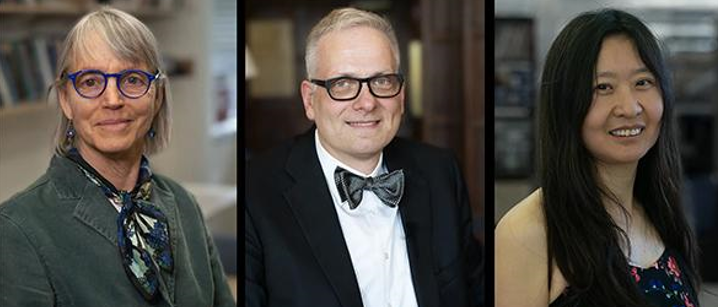
Professors Nancy Kanwisher, Winrich Freiwald and Doris Tsao were jointly awarded the 2024 Kavli Prize for Neuroscience. Adapted from https://www.nei.nih.gov/about/news-and-events/news/2024-kavli-prize-neuroscience-awarded-discovery-face-areas-brain.
The Human Face Area: A Breakthrough in Brain Imaging
The ability to distinguish and identify faces is essential for effective communication and building social bonds, as well as for recognising individuals across different contexts. Yet, the exact neural system responsible for this function had long remained unclear. While earlier research in psychology and animal studies suggested the existence of a specialised mechanism for face recognition, the specific brain regions involved had not been definitively identified.
Prof Nancy Kanwisher (Ref.1) and her team used functional magnetic resonance imaging (fMRI; Ref.2) to scan brain activity as human subjects viewed faces and objects. Their work led to the identification of a small region of the neocortex, now known as the fusiform face area (FFA), which responds selectively to faces (Figure 1; Ref.3). This discovery not only confirmed the existence of a face-specific recognition system in the human brain but also introduced new techniques for identifying specialised brain regions. Prof Kanwisher’s methods for localising functional areas have since become widely used across neuroscience, extending beyond face recognition to other cognitive domains.
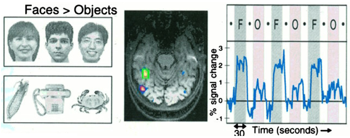
Figure 1. The fusiform face area (FFA), as identified by fMRI, showing stronger activation in response to faces compared to objects. Adapted from Ref. 3.
Extending the Discovery: From Humans to Monkeys
Building on Prof Kanwisher's discoveries, Prof Doris Tsao (Ref. 4) collaborated with Prof Winrich Freiwald to explore whether similar face-specific regions could be found in non-human primates. Through fMRI, they identified six distinct "face patches" in the macaque brain, each specialised for processing different aspects of facial information (Figure 2; Ref. 6). Together, they mapped the functional organisation of these patches, revealing a complex neural network dedicated to face recognition.
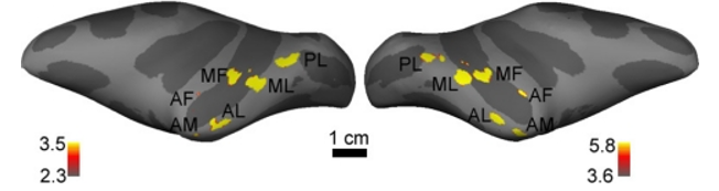
Figure 2. Face-selective regions in the monkey brain, identified through fMRI, shown on a lateral view of the inflated hemispheres. Adapted from Ref. 6.
They further investigated by inserting electrodes into these face patches, recording the activity of individual neurons. They found that many neurons in these areas responded strongly and selectively to faces, while these neurons were almost completely silent in response to non-face images (Figure 3; Ref.7). This fMRI-guided electrophysiology approach provided unprecedented insight into the macaque inferotemporal cortex, showing how groups of neurons collaborate to process facial features.
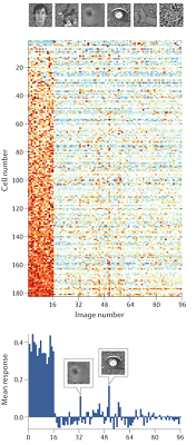
Figure 3. fMRI-guided electrophysiology in a macaque monkey face patch reveals that the majority of neurons exhibit strong responses to faces compared to other images. Adapted from Ref. 7.
Prof Tsao and her team advanced their research by revealing how the face patches work together to recognise faces. They discovered a neural "code" in which different neurons are tuned to specific facial features, such as hair or the distance between the eyes. Impressively, by analysing the activity of just a few hundred neurons, the researchers were able to reconstruct the faces that the monkeys were viewing (Ref. 8). Freiwald later demonstrated that another region, the temporal pole, plays a key role in recognising familiar faces, with certain neurons selectively responding to faces previously encountered (Ref. 9).
A Collaborative Legacy in Neuroscience
Professors Kanwisher, Freiwald, and Tsao’s combined efforts have illuminated the specialised brain machinery dedicated to face recognition, spanning from the discovery of the human fusiform face area to the mapping of face patches in monkeys. Their research not only highlights the brain’s remarkable ability to process social information but also provides broader implications for understanding how we recognise objects and scenes. By elucidating the neural basis of face recognition, they have laid the groundwork for future explorations into the intricate connections between perception, memory, and social cognition.
References
Ref. 1 (Nancy Kanwisher) https://web.mit.edu/bcs/nklab/
Ref. 2 (fMRI) https://en.wikipedia.org/wiki/Functional_magnetic_resonance_imaging
Ref. 3 (FFA) https://pmc.ncbi.nlm.nih.gov/articles/PMC6573547/
Ref. 4 (Doris Tsao) https://tsaolab.berkeley.edu/dortsao/
Ref. 5 (Winrich Freiwald) https://lab.rockefeller.edu/freiwald/
Ref. 6 (Face patches) https://pmc.ncbi.nlm.nih.gov/articles/PMC2614792/
Ref. 7 https://pmc.ncbi.nlm.nih.gov/articles/PMC2678572/
Ref. 8 https://pmc.ncbi.nlm.nih.gov/articles/PMC8088389/
Ref. 9 https://pmc.ncbi.nlm.nih.gov/articles/PMC8645414/