Magnetoencephalography (MEG)

Overview
How does MEG work?
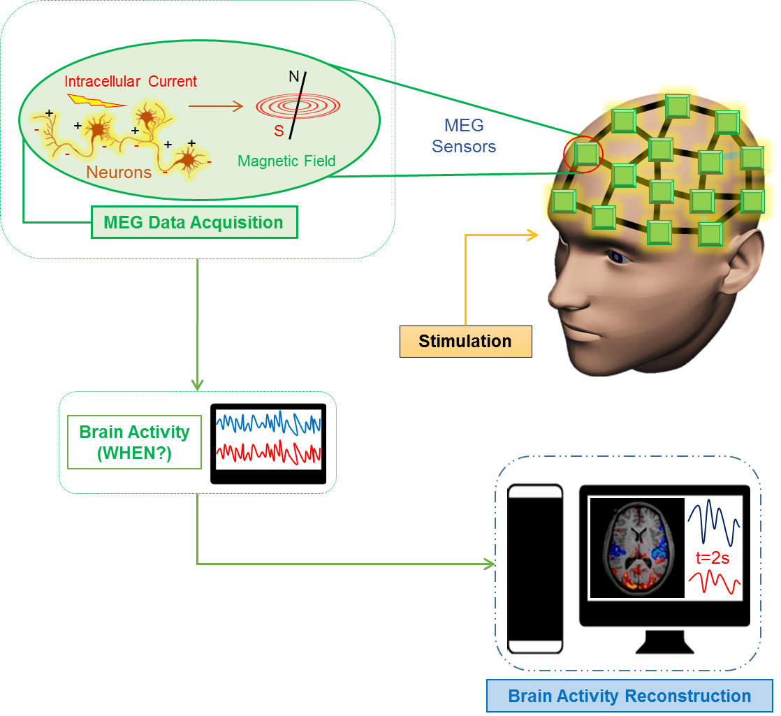
SQUIDs can detect tiny magnetic signals, much less than one-billionth the strength of the Earth’s magnetic field, and then convert these into recordable electric voltages. The SQUID array is mounted in a close-fitting helmet and is cooled with liquid helium. Neuronal signals may be events lasting from about a millisecond for an action potential, tens to hundreds of milliseconds for postsynaptic potentials or even multiple seconds for modulation of brain rhythms.
What is MEG used for ?
Common clinical applications include Detection of Epileptic Activity and Presurgical Functional Mapping. Researchers continue to use MEG to provide new insights into the neural basis of developmental disorders such as autism and dyslexia, as well as psychiatric diseases including depression, bipolar disorder and schizophrenia. Neurodegenerative diseases such as Alzheimer’s disease are also increasingly studied.


Core MEG System
Core MEG System
306 Channel Magnetoencephalography (MEG) System
The core system at CoNiC is a state-of-the-art 306-channel system for MEG measurements (Elekta Neuromag TRIUX) with an integrated 128-channel EEG system (Elekta), located inside a 3-layer magnetically shielded room (Vacuumschmelze GmbH), and related amplifiers and acquisition systems.

Elekta Neuromag® TRIUX employs thin-film sensors of two different types integrated on 102 sensor elements. Each is equipped with three independent sensors with different sensitivity patterns.Each sensor element contains a magnetometer that measures the normal field component. These sensors are highly attuned to all signals, whether from deep or superficial sources - regardless of orientation. Each sensor element also provides two orthogonal planar gradiometer sensors for measuring the gradient components. The primary advantage of this triple-sensor design is that it provides a combination of three unique and mutually independent measurements instead of oversampling the same information as would be the case with an axial gradiometer or magnetometer-only system.
 The lab is uniquely equipped with wide array of best-available equipment stimulation (visual, auditory, somatosensory, olfactory and more), measurement of and registration of responses (psychophysiology, eye tracking, behaviour and more) all handpicked, modified, installed and verified for millisecond timing in the MEG environment.
Integrated 128 Channel EEG
Integrated Bipolar Channels
Triggers and Time-Locking
Magnetically Shielded Room (MSR)
Data Analysis Software
- Signal Processor
- Signal Plotter
- Source modeling
- MRI integration
- Minimum current estimation (for non-diagnostic use only)
Data Storage & Management
The data generated per acquisition in MEG is of Gigabytes. The data generated is locally stored in the aquisition work station, and periodically will be transferred to our Network Attached Storage (NAS). The total capacity of NAS is about 50TB. The total storage has been fragmented to about 25TB for MRI and 20TB for MEG respectively. All the storage and back-up systems are connected by local network to ensure smooth & safe data transfer and storage. The NAS has been configured to be compatible to store the MEG data in the required format. The handling of data and its delivery to the collaborators are followed according to the SOP.

Stimulation Equipment
1. Auditory Stimulation
The MEG system is equipped with a binaural auditory stimulator with two tubal-insert earphone sets for eliciting auditory evoked fields. The auditory stimulation paradigms for the subjects are typically controlled via Compumedics Neuroscan's STIM2 stimulus presentation and experimental design system.
STIM2 is fully integrated with the Cedrus StimTracker system which uses photocells and auditory threshold detection to identify stimulus onset with the greatest accuracy possible.
2. Somatosensory Stimulation
The MEG system is also equipped with an electrical stimulator for eliciting somatosensory evoked fields. It consist of Digitimer's DS7A single channel current stimulator system and a stimulus electrode with felt tips for median nerve stimulation.

Response Equipment
Finger Response System. The MEG system is equipped with a bilateral finger response system working on fiber optics for detecting subject responses.
Processes
MEG Workflow
Workflow for Subject Preparation & MEG Measurement

Cognitive Tasks
MEG system can provide real-time mapping of brain activity by non-invasively measuring the magnetic fields produced by the brain. The MEG system @ CoNiC is basically focused on studying cognitiion based tasks which includes basic research within cognitive neuroscience in the areas of memory, language, attention, perception, pain, music, decision making, emotion and others, performed on healthy adult populations.
The STIM2 system with our MEG is a comprehensive stimulus presentation system consisting of a library of sensory, cognitive and neuropsychological tasks. It contains well-defined and widely known paradigms for cognitive tasks.
STIM2 is fully integrated with the Cedrus Stim Tracker. This system uses photocells and auditory threshold detection to identify stimulus onset with the greatest accuracy possible.
Principle Categories of Tasks
Fourteen tasks are pre-programmed into the STIM2 software to provide a task library to build upon. Each task allows the user to modify parameters, such as, the duration, order of presentation, the inter-stimulus interval, performance feedback options, and many more. The programs are categorized as follow:

Additional tasks include pattern reversal, Naming, Visual tracking, spatial memory, Visual and Auditory continuous performance, Verbal learning, and Visual memory tasks.
MEG Safety
MEG: A Safe Environment
A MEG scan is noninvasive and painless with no injections, radioactivity or strong magnetic fields. MEGs have highly sensitive SQUID Sensors which needs to be protected from external magnetic fields and other noises.
Getting Screened for Scan

Precautions for Entering MEG Scanning Room

Safety Incharge


/enri-thumbnails/careeropportunities1f0caf1c-a12d-479c-be7c-3c04e085c617.tmb-mega-menu.jpg?Culture=en&sfvrsn=d7261e3b_1)

/cradle-thumbnails/research-capabilities1516d0ba63aa44f0b4ee77a8c05263b2.tmb-mega-menu.jpg?Culture=en&sfvrsn=1bc94f8_1)

