Biomolecular Interactions
The Biomolecular Interactions (BMI) platform in School of Biological Sciences at NTU offers access to a range of different biophysical instruments to NTU community and external users. The facility houses different instruments that can help users characterize molecules of their research and interest. We time and now have pursued to expand our facility and we hope to incorporate more resources in future that can help advance science.
The BMI facility is open to NTU, other academic as well as Industrial researchers.
Please find more information on the currently available equipment and their scope below.
CONTACT INFORMATION
| Location of Facility | School of Biological Sciences SBS-04S-43 |
| Contact Details | Devanshu Mehta, Ph.D. In-charge, Biomolecular Interaction (BMI) Facility Email Address: Devanshu.mehta@ntu.edu.sg |
SURFACE PLASMON RESONANCE
 DESCRIPTION
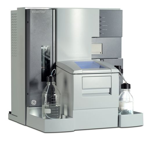
We have the state-of-the-art Biacore T200 SPR available.
The SPR technology in Biacore systems is used to monitor binding events between molecules ranging from ions to viruses. The technology provides binding kinetics (ka and kd), affinity (KD), specificity and concentration, without any needs for labels.
This technology can help you characterize interactions between:
- Protein to Protein
- Protein to DNA
- Protein to Sugar
- Protein to small molecular / drug / fragments / hormones
- Antibody interactions
- Affimers
- And many more! As long as the binding partners are soluble and there are means to couple one of them onto a sensor chip surface.
Through this technique, you can determine the strength of interactions, as well as the real time kinetics, i.e. the association and dissociation rates, which can allow you to reach new research conclusions.
In the Biacore T200, the built-in microfluidic system creates on the surface of a sensor chip four flow cell channels in serial flow design. The first flow cell is commonly used as a reference to subtract out refractive index difference and non-specific bindings. For typical binding experiments, active referencing is achieved when two channels are used and samples are serially injected across the reference flow cell followed by the ligand flow cell. The sensorgrams obtained from the reference and active flow cells are then subtracted to get specific binding curves and kinetic parameters of the interaction studied.
SPR AT NTU
In 2007, an SPR Biosensing platform was established in Singapore by Associate Professor Susana Geifman at the School of Biological Sciences, NTU, serving the SPR biosensing needs of NTU and other institutions. Testimony to the importance and success of the technology, the SPR Biosensing platform had successfully carried out hundreds of collaborations, projects and contributed to many publications. Data from SPR had also help PIs strategize and refine their research finding/directions.
In 2009, Professor Bo Liedberg from Linköping University, Sweden, came to NTU on a Visiting Professorship and joined NTU as a full faculty in 2012. He established the Centre for Biomimetic Sensor Science in NTU, together with the SPR platform at SBS, many high quality, international molecular interaction workshops and events had been organized, such as the Advanced Biosensor Workshop in 2011, the 30 Years of SPR Biosensing symposium in 2013, etc.
For a quick introduction on Biacore T200 SPR, read here
BIOLAYER INTERFEROMETRY
 |  |
DESCRIPTION
We have next-gen Gator Prime Biolayer interferometer available.
Biolayer interferometry (BLI) is a label-free, fluidic-free, biomolecular interaction detection method. The technique measures difference in white light interference at a biosensor surface to detect binding interactions in real time. Gator prime offers user-friendly, high-throughput, and quick data acquisition for kinetic, quantitation and epitope binning experiments. Gator prime is an 8-channel system with two-96-well plate capacity and ability to control measurement temperature from ambient to 40degC. The machine offers detection of binding affinities in the range of 10pM – 1mM. The technique can be used for proteins, antibodies, peptides, nucleic acids, liposomes, viruses, small molecules (>150 Da), and more.
This technology can help you:
- get information on binding kinetics (rates of association, ka, and dissociation, kd), affinity (KD), specificity, and concentration.
- Save precious samples, sample retrieval post-experiment.
- Run eight different samples in the same cycle.
- Run purified or crude samples.
- Analyze biological molecules at different stages of therapeutic development.
- Provide information on yes/no binding.
- Assess antibody affinity maturation, activity, and stability; and more!
For a quick introduction on details for setting a kinetic experiment on Gator Prime Biolayer interferometer, download the pdf here.
ISOTHERMAL TITRATION CALORIMETRY

DESCRIPTION
We have Malvern iTC200 available.
Isothermal Titration Calorimetry (ITC) is used to measure reactions between biomolecules. The methodology allows quantitative determination of the binding affinity, stoichiometry, entropy, and enthalpy of the binding reaction in solution, without the need to use labels or to immobilize molecules.
When binding occurs, heat is either absorbed or released and this is measured by the sensitive calorimeter during gradual titration of the ligand into the sample cell containing the biomolecule of interest.
This technology can help you in:
- Quantification of binding affinity
- Candidate selection and optimization
- Measurement of thermodynamics and active concentration
- Characterization of mechanism of action
- Confirmation of intended binding targets in small molecule drug discovery
- Determination of binding specificity and stoichiometry
- Validation of IC50 and EC50 values during hit-to-lead
- Measurement of enzyme kinetics
The ITC200 requires approx 240ul of Interactant A, and 40ul of Interactant B.
The selection criteria are on solubility, and on which interactant is easier to obtain homogenously and stably at higher concentrations. Recommended concentrations can range between 3uM to 0.5mM.
MASS PHOTOMETERY
 |  |
DESCRIPTION
We have Refeyn TwoMP Mass photometer available.
Mass photometer is a label-free, highly sensitive, molecular mass measurement method. The technique requires only minimal sample quantity (few microlitres), 5-20nM sample concentration to perform mass analysis in minutes. Refeyn TwoMP can efficiently measure samples ranging in size from 30kDa to 5MDa. It has a user-friendly, quick data acquisition interface for molecular mass measurements. The technique can be used for proteins, DNA, RNA, large macromolecular complexes, nanostructures, and even small viruses such as AAVs, and more.
This technology can help you:
- Measure accurate molecular mass of your samples.
- Measure at physiological (i.e. low) concentrations.
- Capture low-abundance species.
- Study protein-protein, protein-DNA interaction, complex formation, and protein oligomerisation.
- Assesse sample heterogeneity, purity, and stability prior to biochemical and structural studies.
- Calculate equilibrium dissociation constant (KD) for biomolecular interactions; and more!
For a quick introduction on Refeyn TwoMP Mass photometer, read here
DYNAMIC LIGHT SCATTERING

DESCRIPTION
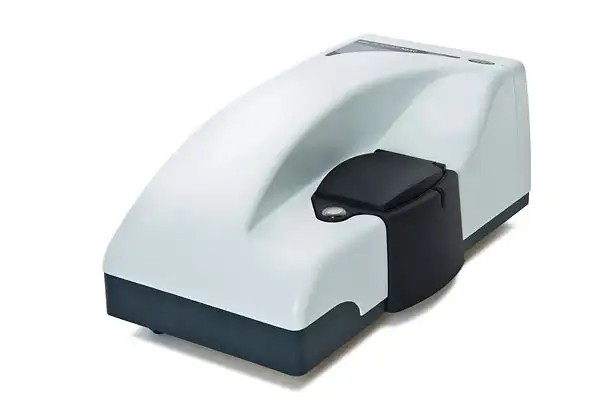
We have Malvern Zetasizer ZS-Nano available.
The Zetasizer Nano ZS is a very quick, and extremely useful instrument to learn and use. The NanoZS model allows us to size molecules of diameter between 0.3nm and 10 microns. Whether it is proteins, or bacterial cells in solution, the NanoZS allows users to rapidly obtain information on the population distribution of sizes within a sample, with sample being recoverable. Typical disposable cuvettes require approx 400ul of sample.
This technology can help you:
- determine protein sample homogeneity,
- perform oligomerization studies,
- test sample quality over time,
- Use it for protein "melting" with its heating function up to over 95 degrees celsius.
- Measure Zeta potentials, the velocity/speed of particles in solution when subjected to an electric field can also be measured on this instrument.
CIRCULAR DICHROISM

DESCRIPTION
We have Applied Photophysics Chirascan Circular Dichroism Spectrometer available.
Circular Dichroism is the differential absorption between left and right circularly polarized light by chiral molecules or environment by a sample.
This technology can help you characterize:
- Changes in secondary and tertiary structure of samples (proteins, RNA etc).
- Quantify secondary structure content of proteins (like % of alpha helices, beta sheets, random coils etc.).
- Effect of mutations on conformation of wild type proteins.
- Batch to batch consistency in quality of sample preparation.
- Sample stability as a function of time and temperature.
- Conformational changes to samples in presence of ligands or drugs; and many more!
For a quick introduction on Circular Dichorism by Applied Photophysics, download the pdf here
FLUORIMETERY
 DESCRIPTION
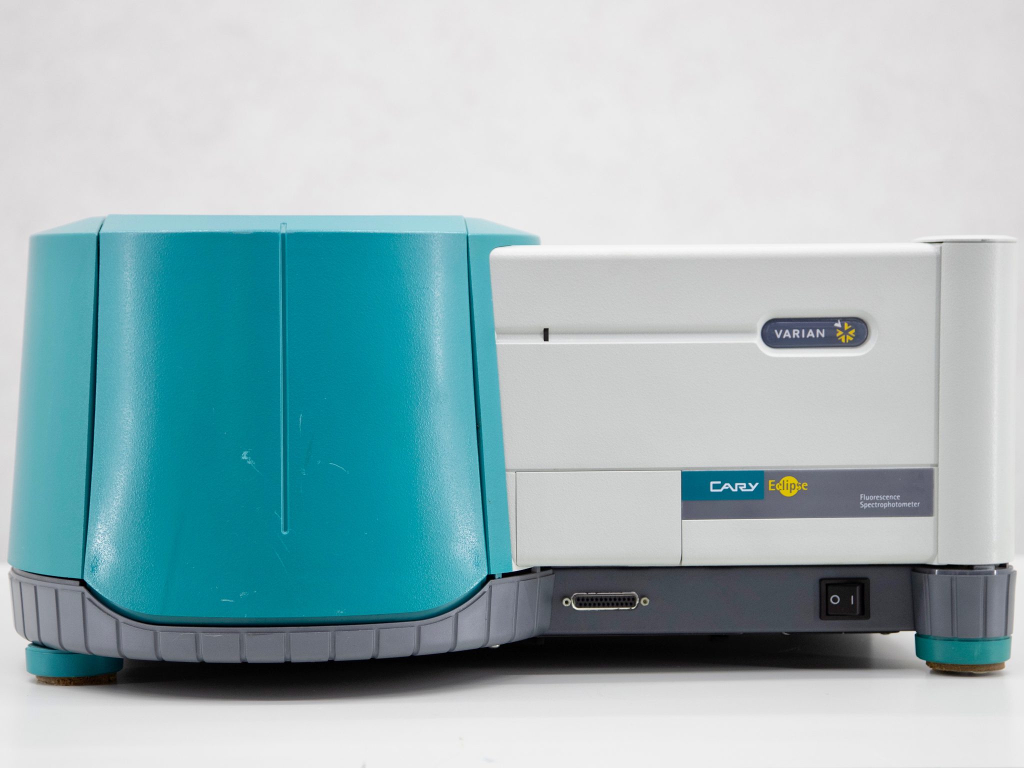
We have Agilent Varian Cary Eclipse Fluorescence Spectrometer available.
Fluorescence Spectrometer can be used to record fluorescence, phosphorescence, chemiluminescence, and bioluminescence measurements. The fluorometer has a wavelength range of 200 – 900 nm for both excitation and emission monochromators.
This technology can help you:
- Perform kinetic measurements.
- Measure changes to Intrinsic fluorescence of proteins.
- Study protein stability
- Study and identify protein aggregation; and more.
ANALYTICAL ULTRACENTRIFUGATION

DESCRIPTION
We have Beckman ProteomeLab XL-1 Analytical Ultracentrifuge (AUC) available.
Analytical ultracentrifugation is a high-speed centrifuge technique that can help monitor the behaviour of particles in real time. ProteomeLab XL-I can help characterize a protein’s behaviour in free solution under native conditions. The system can work under a variety of conditions of concentration, temperature, ionic strength and pH and help in thorough characterisation of molecules. The technique can help provide information about the size and shape of molecules and also of solution molar masses, stoichiometries, association constants and solution nonideality.
This technology can help you:
- Characterize sample heterogeneity for process development, formulation and QA/QC.
- Determine yes/no binding of a small molecule to a target protein.
- Characterize molecular conformation under a variety of conditions.
- Characterise molecular weight, stoichiometry, aggregation, self-association, conjugation efficiency, polydispersity and more for proteins and complexes.
For a quick introduction on Analytical Ultracentrifugation by Beckman Coulter, download the pdf here
BMI Facility charges
Equipment | Cost Scheme | ||
NTU | Academic Institutions | Industry | |
Biacore T200 SPR (Assisted) | S$250/day + chip(s) | S$330/day + chip(s) | S$415/day + chip(s) |
Gator Prime BLI | S$60/hour + consumables | S$100/hour + consumables | S$120/hour + consumables |
Malvern ITC200 | S$215/day | S$290/day | S$360/day |
Malvern Zetasizer ZS Nano | S$20/hour | S$50/hour | S$60/hour |
Refeyn TwoMP Mass photometer | S$80/hour + consumables | S$120/hour + consumables | S$140/hour + consumables |
Chirascan CD Spectrophotometer | S$40/hour | S$80/hour | S$100/hour |
Cary Eclipse Fluorometer | S$30/hour | S$50/hour | S$60/hour |
Analytical UC | S$77/hour (trained user) | S$250/day +Assistance | S$300/day + Assistance |
Note:
| |||
Access and usage policy of BMI platform
- Training and Assistance: Interested new users can request training for machines under BMI facility at an hourly chargeable rate. Training sessions do not include data collection. During training, the users are informed on the modus
operandi of machine hardware/software, experimental flow and maintenance. Assistance to troubleshoot/optimize/analyse can also be requested by users from BMI staff at an hourly chargeable rate.
- When working in facility: Always keep the BMI staff informed on the number of people working on the instrument. If you don’t have access to the facility or machine booking on CEBS, please contact BRC at brc@ntu.edu.sg. Users must follow proper SOP and should contact the BMI staff in case of any problem or doubt. The users must use appropriate PPE. Users must appropriately fill required logbooks to facilitate proper
dispense of information on usage and billing. Users must back-up all data; BMI has no responsibility on data loss.
- During publications: Users are requested to acknowledge the support of Biomolecular Interaction facility, NTU in their publications. Proper acknowledgement of BMI’s contribution to your published work helps us in evaluations and proper funding. Further, in case of substantial and important intellectual contribution by BMI staff member(s), a proper consideration for inclusion in authorship as accepted in typical academic collaborations is highly appreciated. The users can share their publication information to sbs-bmi@ntu.edu.sg to help us document our support in your scientific journey.


/enri-thumbnails/careeropportunities1f0caf1c-a12d-479c-be7c-3c04e085c617.tmb-mega-menu.jpg?Culture=en&sfvrsn=d7261e3b_1)

/cradle-thumbnails/research-capabilities1516d0ba63aa44f0b4ee77a8c05263b2.tmb-mega-menu.jpg?Culture=en&sfvrsn=1bc94f8_1)
